Biomanufacturing
Development of a Robust Human-Mouse Hybrid in vitro Model of Acute Respiratory Distress Syndrome for Stromal Cell Therapy
(C-89) Development of a Robust Human-Mouse Hybrid in vitro Model of Acute Respiratory Distress Syndrome for Stromal Cell Therapy

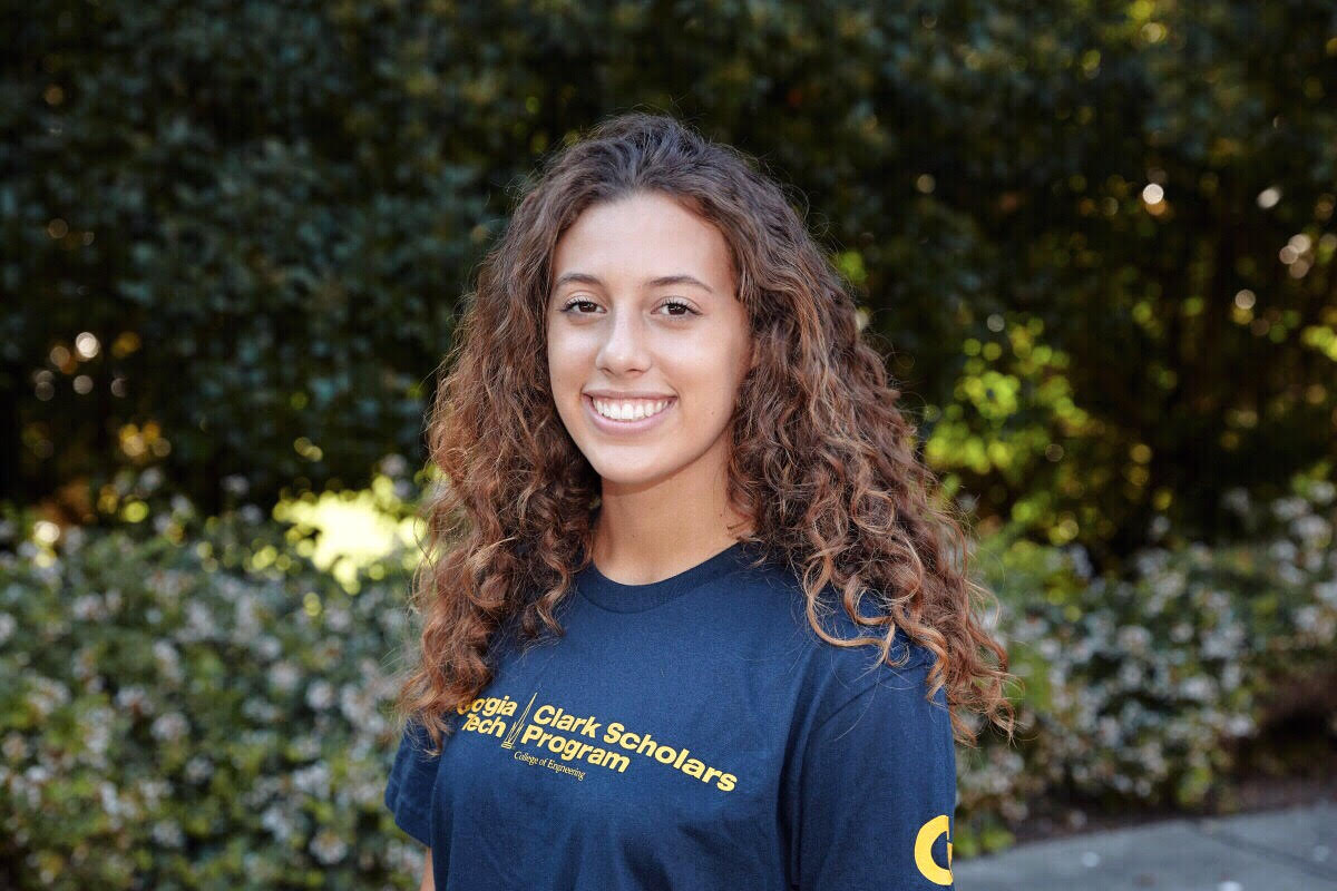
Nicole Frey (she/her/hers)
Undergraduate Student
Georgia Institute of Technology
Cartersville, Georgia, United States
Angela Jimenez, MS (she/her/hers)
Doctoral Student
Georgia Institute of Technology
Orlando, Florida, United States- KR
Krishnendu Roy
Professor
Georgia Institute of Technology
Atlanta, Georgia, United States
Presenting Author(s)
Co-Author(s)
Primary Investigator(s)
Acute Respiratory Distress Syndrome (ARDS) is a complex and life-threatening condition characterized by severe lung injury, resulting in high mortality rates (40%) worldwide.1 The detrimental consequences of ARDS are multifaceted, beginning with impaired gas exchange in the alveoli, leading to systemic inflammation and multiple organ dysfunction syndrome.2 These critical complications compromise patient outcomes and impose an immense burden on healthcare resources.2 Despite extensive research efforts, the precise pathophysiological mechanisms underlying ARDS are not yet fully known, posing substantial challenges to the development of effective therapeutic interventions.2 As a result, there are no therapeutics available to patients, only strategies to mitigate pain and discomfort. Stem/stromal cell therapies show promise in treating various diseases and injuries by facilitating tissue repair, cellular restoration, and immune modulation.3 Mesenchymal Stromal Cells (MSCs), found in bone marrow, adipose tissue, umbilical cord, and connective tissues, offer therapeutic benefits through potent immunomodulation and anti-inflammatory properties.3 In the context of ARDS, where a large influx of immune cells including macrophages and neutrophils contribute to lung tissue damage, MSCs have shown the ability to reduce inflammation and tissue damage in the lung environment.4 These properties position MSCs as a potential therapeutic option for addressing inflammation-related lung diseases like ARDS. As a result, in this study, we aimed to develop a human-mouse hybrid in vitro model of ARDS to investigate the therapeutic potential of MSCs in reducing inflammation and promoting lung repair.
Materials and Methods::
An in vitro model system mimicking an alveolar sac was designed and robustly examined using a transwell setup. To mimic the physiological structure of the lung microenvironment, primary mouse lung epithelial cells were cultured on the apical side of the transwell membrane, while primary mouse lung endothelial cells were seeded on the basolateral side. Various combinations of seeding densities, extracellular attachment proteins, and time points were examined to ensure uniform monolayer formation and presence throughout the duration of the assay. Transepithelial electrical resistance (TEER) measurements and confocal microscopy were performed to evaluate cellular integrity and barrier function. THP-1s, a human monocytic cell line, were added to the top chamber of the transwell after media formulation testing, and validated activation and polarization responses. A solution of lipopolysaccharides (LPS) was used to induce cellular injury and inflammation as presented in ARDS. To ensure a heightened response, various concentrations and time points of LPS exposure were optimized and assessed with flow cytometry for toll-like receptor presence and Luminex analysis to assess the inflammatory response and proinflammatory cytokine production. After LPS exposure, MSCs were administered in the bottom endothelial layer and the reparative response was measured with various quantitative outputs. Media from the apical side was collected for Luminex analysis and a permeability assay using FITC-Dextran was performed to evaluate cellular integrity and barrier function. Flow cytometry was employed on the THP-1s to confirm the presence of M1-like markers in response to LPS and to determine the macrophage polarization state when MSCs were present.
Results, Conclusions, and Discussions::
To develop the human-mouse hybrid in vitro ARDS model, optimization of all cellular types and quantitative outputs, workflow, and time points was completed. Through continuous TEER measurements and confocal microscopy, we successfully employed different seeding densities across the cell types to show the robust model over a 10-day assay. LPS experiments aimed to optimize the desired concentration and incubation time required to induce an inflammatory response representative of ARDS. The results of the permeability assay showed increased monolayer permeability with longer LPS incubation times, indicating damage to tight junction barriers and toll-like receptor activation was confirmed via flow cytometry. Incorporating THP-1-derived naive macrophages into the in vitro system required preliminary testing to understand how different media conditions could affect cell viability and activation. The THP-1s were cultured in various media combinations and after incubation, showed no visible impact on cell viability, growth, or morphology, indicating suitability for further experiments. Macrophage activation/polarization assays confirmed that THP-1s exposed to LPS polarized into the M-1 phenotype, as expected, regardless of the media used. When MSCs were added to the system over 72 hours, we observed M2-like phenotype changes indicating MSCs are reducing the inflammatory state and promoting repair. Given the complexity of the human-mouse hybrid system, optimizing the in vitro model for the integration of both human and mouse cell lines was crucial for the model’s development. Through this work, we examined the incorporation of each competent of the model and optimized the model parameters so that it has the potential to accurately predict the therapeutic effects of MSCs in an in vivo humanized mouse.
Acknowledgements (Optional): :
References (Optional): :
< ![if !supportLists] >1. < ![endif] >Ramji HF, Hafiz M, Altaq HH, Hussain ST, Chaudry F. Acute Respiratory Distress Syndrome; A Review of Recent Updates and a Glance into the Future. Diagnostics (Basel). 2023 Apr 24;13(9):1528. doi: 10.3390/diagnostics13091528. PMID: 37174920; PMCID: PMC10177247.
< ![if !supportLists] >2. < ![endif] >Diamond M, Peniston HL, Sanghavi DK, et al. Acute Respiratory Distress Syndrome. [Updated 2023 Apr 6]. In: StatPearls [Internet]. Treasure Island (FL): StatPearls Publishing; 2023 Jan-. Available from: https://www.ncbi.nlm.nih.gov/books/NBK436002/
< ![if !supportLists] >3. < ![endif] >Coelho A, Alvites RD, Branquinho MV, Guerreiro SG, Maurício AC. Mesenchymal Stem Cells (MSCs) as a Potential Therapeutic Strategy in COVID-19 Patients: Literature Research. Front Cell Dev Biol. 2020 Nov 19;8:602647. doi: 10.3389/fcell.2020.602647. PMID: 33330498; PMCID: PMC7710935.
< ![if !supportLists] >4. < ![endif] >Zhang, X., Wei, X., Deng, Y. et al. Mesenchymal stromal cells alleviate acute respiratory distress syndrome through the cholinergic anti-inflammatory pathway. Sig Transduct Target Ther 7, 307 (2022). https://doi.org/10.1038/s41392-022-01124-6
