Cancer Technologies
(F-230) OPEN-SOURCE ROBOTIC PLATFORM FOR AUTOMATED MICROMANIPULATION OF LIVE MICROTISSUES

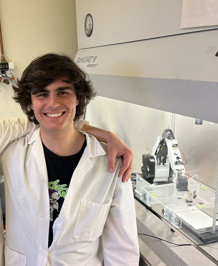
Noah Gottshall (he/him/his)
Undergraduate research assistant
University of Washington Seattle
Normandy Park, Washington, United States- AF
Albert Folch (he/him/his)
Professor of Bioengineering
University of Washington, Washington, United States - DS
Daksh Sinha
Research assistant
University of Washington Seattle, United States - IS
Ivan Stepanov
Research Assistant
University of Washington Seattle, United States - SH
Sarmad Hassan, n/a
Research assistant
University of Washington, United States - SB
SangYoon Back
Research assistant
University of Washington Seattle, United States
Presenting Author(s)
Primary Investigator(s)
Co-Author(s)
Human donor tissue is generally scarce. Hence there has been a rising interest in the production of submillimeter-sized 3D tissues (“microtissues”), either bioprinted, (1) microengineered, (2,3) or aggregated from single cells (4,5) or, conversely, microdissected from larger biopsies (6-13) (encompassing organoids, organospheres, spheroids, tumoroids, cuboids, etc.). Microtissues are increasingly used for disease modeling in cancer (14) and immunology, (15) as well as for regenerative medicine, (16) personalized medicine, (17-19) and drug screening. (17,20) To broaden the access to microtissue research, we have developed an open-source platform that can be programmed to quickly size-sort and pick the microtissues one by one from a random distribution of microtissues (e.g. in a petri dish) and to place them into a 96-well plate with the help of an inexpensive robotic arm. We then demonstrate drug testing on cancer microtissues.
Materials and Methods::
The robotic platform utilizes the Dobot MG400 robot, with XYZ accuracy of 50-100 µm, a rotational “head” motor, Python programmability, compact size (fits in a culture hood), and low cost. We use OpenCV computer vision techniques to locate cuboidal-shaped microdissected U87 mouse xenograft glioma tumor cuboids (average ~(400 µm)3 volume), prepared using a tissue chopper (21). The robotic arm takes instructions via Python using visual information gathered from the USB camera.
Computer vision. Using the camera data, a linear transformation matrix is used to create a synchronized coordinate system between the plane of the petri dish and the robotic movement grid. Cuboids are located using light-adaptive thresholding techniques to distinguish cuboids sizes, and relative positions. The highest pixel area frequency range is displayed on a histogram to determine if a cuboid is transferred to the 96-well plate.
Custom pump. We custom-designed and built a peristaltic ring bearing pump that attaches to the stepper motor on the robot’s head. The pump displaces 10 μL per 90 degrees of motor rotation. A glass capillary tube (ID 1 mm) is used for cuboid manipulation.
Drug treatment. Cuboids in the approximate size of (400 µm)3 are transferred to a 96 well plate, from a culture dish containing growth medium. Six different drugs are added in low and high concentrations to desired sections of the plate. Two control columns are used to compare the drug treatments. After 3 days of incubation, the full 96 well plate can be stained and analyzed under a fluorescent microscope.
Results, Conclusions, and Discussions::
In our robotic platform (Fig. 1), a USB camera atop the petri dish size-sorts the microtissues (Fig. 1A) and feeds the position of the microtissues to the robot’s software sequentially (Fig.1B). The platform fits into the culture hood for sterile workflows (Fig. 1C). We integrated a custom-made rotary pump with the head’s motor (Fig. 1D-F) to generate the fluid motion needed to pick and place microtissues.
We used the platform to pick cuboids from mouse tumors (Fig. 2A). A theoretical fluidic analysis illustrates why our setup does not require high-precision instrumentation to position the pipette in z (Fig. 2B,C). We determined that the suctioning system was effective at picking a given cuboid if the pipette mouth was at a height z between 1.0-1.6 mm above the cuboid (Fig. 2D). The setup can also adapt to other applications with smaller and larger microtissues simply by changing the size threshold (Fig. 3A-D). Desired microtissue size ranges can be selected in order to ignore debris and collect more uniform samples (Fig. 3A-D).
Robotic loading facilitates straightforward drug testing in multi-well plates with fluorescent readouts (Fig. 4A-D), which could open new horizons and collaborations in drug testing in a variety of fields. Experiments with live mouse tumors suggest no significant interference in the viability of the microtissues, demonstrating that drug testing is possible with the robotic platform (Fig. 4D). Crucially, the platform is agnostic to the type of tissue being used. Therefore, the platform could easily be extended to human tumor cuboids to allow for tests that are relevant to the human TME and to inform drug testing modalities such as immunotherapy that require the preservation of the TME in vitro.
Acknowledgements (Optional): :
References (Optional): :
REFERENCES
1. Banerjee, D., Singh, Y. P., Datta, P., Ozbolat, V., O’Donnell, A., Yeo, M. & Ozbolat, I. T. Strategies for 3D bioprinting of spheroids: A comprehensive review. Biomaterials 291: 121881 (2022).
2. Leong, T. G., Randall, C. L., Benson, B. R., Zarafshar, A. M. & Gracias, D. H. Self-loading
lithographically structured microcontainers: 3D patterned, mobile microwells. Lab Chip 8: 1621–1624 (2008).
3. Wang, Y., Phillips, C., Xu, W., Pai, J. H., Dhopeshwarkar, R., Sims, C. E. & Allbritton, N. Micromolded arrays for separation of adherent cells. Lab Chip 10: 2917–2924 (2010).
4. Moscona, A. & Moscona, H. The dissociation and aggregation of cells from organ rudiments of the early chick embryo. J. Anat. 86: 287–301 (1952).
5. Gunti, S., Hoke, A. T. K., Vu, K. P. & London, N. R. Organoid and spheroid tumor models: Techniques and applications. Cancers (Basel). 13: 1–18 (2021).
6. Fjellbirkeland, L., Laerum, O. D. & Bjerkvig, R. Tumour fragment spheroids from human non-small-cell lung cancer maintained in organ culture. Virchows Arch. 426: 169–178 (1995).
7. Lao, Z., Kelly, C. J., Yang, X. Y., Jenkins, W. T., Toorens, E., Ganguly, T., Evans, S. M. & Koch, C. J. Improved methods to generate spheroid cultures from tumor cells, tumor cells & fibroblasts or tumor-fragments: microenvironment, microvesicles and MiRNA. PLoS One 10: e0133895 (2015).
8. Aref, A. R., Huang, R. Y.-J., Yu, W., Chua, K.-N., Sun, W., Tu, T.-Y., Bai, J., Sim, W.-J., Zervantonakis, I. K., Thiery, J. P. & Kamm, R. D. Screening therapeutic EMT blocking agents in a three-dimensional microenvironment. Integr. Biol. 5: 381–389 (2013).
9. Aref, A. R. et al. 3D microfluidic ex vivo culture of organotypic tumor spheroids to model immune checkpoint blockade. Lab Chip 18: 3129–3143 (2018).
10. Jenkins, R. W. et al. Ex vivo profiling of PD-1 blockade using organotypic tumor spheroids. Cancer Discov. 8: 196–215 (2018).
11. Ootani, A., Li, X., Sangiorgi, E., Ho, Q. T., Ueno, H., Toda, S., Sugihara, H., Fujimoto, K., Weissman, I.L., Capecchi, M. R. & Kuo, C. J. Sustained in vitro intestinal epithelial culture within a Wnt-dependent stem cell niche. Nat. Med. 15: 701–706 (2009).
12. Li, X. et al. Oncogenic transformation of diverse gastrointestinal tissues in primary organoid culture. Nat. Med. 20: 769–777 (2014).
13. Neal, J. T. et al. Organoid Modeling of the Tumor Immune Microenvironment. Cell 175: 1972-1988.e16 (2018).
14. Kretzschmar, K. Cancer research using organoid technology. J. Mol. Med. 99: 501–515 (2021).
15. Bar-Ephraim, Y. E., Kretzschmar, K. & Clevers, H. Organoids in immunological research. Nat. Rev. Immunol. 20: 279–293 (2019).
16. Skylar-Scott, M. A., Uzel, S. G. M., Nam, L. L., Ahrens, J. H., Truby, R. L., Damaraju, S. & Lewis, J. A.Biomanufacturing of organ-specific tissues with high cellular density and embedded vascular channels. Sci. Adv. 5: eaaw2459 (2019).
17. Sachs, N. et al. A Living Biobank of Breast Cancer Organoids Captures Disease Heterogeneity. Cell 172: 373-386.e10 (2018).
18. Pauli, C. et al. Personalized in vitro and in vivo cancer models to guide precision medicine. Cancer Discov. 7: 462–477 (2017).
19. Ding, S. et al. Patient-derived micro-organospheres enable clinical precision oncology. Cell Stem Cell 29: 905-917.e6 (2022).
20. Driehuis, E., Kretzschmar, K. & Clevers, H. Establishment of patient-derived cancer organoids for drug-screening applications. Nat. Protoc. 15: 3380–3409 (2020).
21. Horowitz, L. F., Rodriguez, A. D., Au-Yeung, A., Bishop, K. W., Barner, L. A., Mishra, G., Raman, A., Delgado, P., Liu, J. T. C., Gujral, T. S., Mehrabi, M., Yang, M., Pierce, R. H. & Folch, A. Microdissected “cuboids” for microfluidic drug testing of intact tissues. Lab Chip 21: 122 (2021).
