Cardiovascular Engineering
Human Aortic Valve Interstitial Cell Culture on Valve Mimetic Bilayered Hydrogels to Assess Metabolic and Proteomic Changes during CAVD Progression
(G-280) Human Aortic Valve Interstitial Cell Culture on Valve Mimetic Bilayered Hydrogels to Assess Metabolic and Proteomic Changes during CAVD Progression
- CA
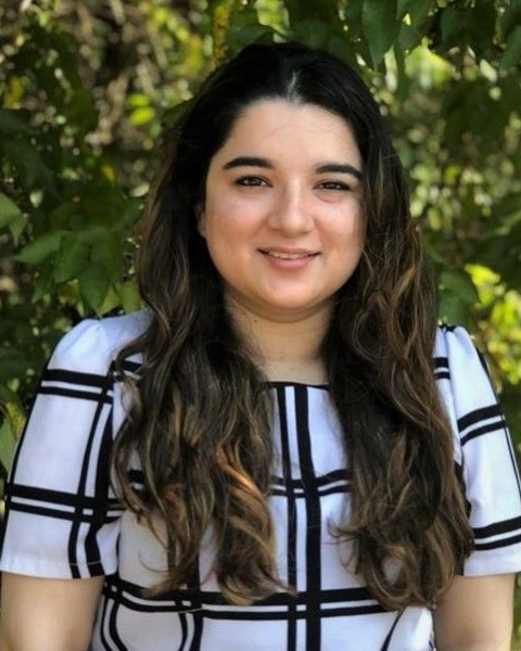
Camryn Adamson (she/her/hers)
REU Student
University of Arkansas, United States 
Ishita Tandon, PhD (she/her/hers)
Postdoctoral Fellow
University of Arkansas
Fayetteville, Arkansas, United States- KB
Kartik Balachandran, PhD
Associate Professor
University of Arkansas
Fayetteville, Arkansas, United States
Presenting Author(s)
Co-Author(s)
Last Author(s)
Calcific Aortic Valve Disease (CAVD) is an active, progressive, disease that affects a wide variety of individuals and is the third leading cause of cardiovascular disorders [1]. CAVD has been shown to initiate aortic stenosis, which can lead to decreased blood flow and ultimately heart failure [2]. Human Aortic Valve Interstitial Cells (HAVIC) play a key role in this dynamic process by maintaining the structural integrity and regulating homeostasis of the aortic valve [3]. Disruption in this homeostasis can occur through osteogenic differentiation of HAVICs, potentiating maladaptive biomechanics [1]. Limited research on CAVD’s progression inhibits effective medical treatments to regulate its adverse effects [2]. In this study, healthy and osteogenic HAVIC cultures were monitored via proteomic and multiphoton assessment in a valve mimetic hydrogel. Based on our preliminary work in porcine VICs [4], we hypothesized that HAVICs, when subjected to an osteogenic three-dimensional environment, will have dysregulated cell metabolism, protein homeostasis, and cholesterol biosynthesis.
Materials and Methods::
HAVICs (Cat. #00225974, Lonza) were maintained in mesenchymal stem cell growth medium (Cat. #PT-4105, Lonza). Hydrogels consisting of a spongiosa- and fibrosa- (two aortic valve layers) mimicking bilayer were created with differing concentrations of collagen1, hyaluronic acid, and chondroitin sulfate, to simulate the healthy and diseased HAVIC microenvironment. Polydopamine treatment was used to adhere the hydrogels to the 48-well plate and cultured over a 21-day period. Regular media was added to the healthy cells and osteogenic media consisting of regular media supplemented with dexamethasone, β-glycerophosphate, ascorbic acid, and transforming growth factor β1 was added to the diseased cells. On day 2, MTS assessment, Alizarin Red staining, and immunocytochemistry were carried out to assess cell viability, calcification, and phenotype, respectively. Primary antibodies against ɑ-SMA and osteopontin were used for phenotypic assessment. Multiphoton microscopy and MTS assay will be performed on days 1, 2, 7, 14, and 21. On days 2 and 21, cell lysates will be prepared for protein sequencing post multiphoton imaging.
Results, Conclusions, and Discussions::
Both healthy and diseased cell types maintained their morphology during seeding on the hydrogels. After 2 days of the cells being placed in healthy and diseased environments, we observed that diseased cells were more elongated, had higher calcification, and increased cell viability. Based on our preliminary data (not published), we also expect dysregulated proteostasis and cholesterol biosynthesis at the early time points. Cells are expected to prefer oxidative phosphorylation to meet their metabolic demands during the disease initiation phase and then switch to glycolysis as the osteogenic differentiation progresses [5].
This study investigates CAVD progression and its effect on HAVICs by utilizing fibrosa and spongiosa mimicking hydrogels. Bilayered hydrogel closely mimicked the healthy and diseased compositions of the aortic valve layers. The diseased bilayered gel composition along with osteogenic media promoted elongated cells with higher viability and increased calcification by day 2. Further assessment will be conducted to reveal changes in metabolic and proteomic profile of healthy and diseased HAVICs during CAVD progression.
Acknowledgements (Optional): :
References (Optional): :
- Duan, B., Yin, Z., Hockaday Kang, L., Magin, R. L., & Butcher, J. T. (2016). Active tissue stiffness modulation controls valve interstitial cell phenotype and osteogenic potential in 3D culture. Acta Biomaterialia, 36, 42–54. https://doi.org/10.1016/j.actbio.2016.03.007
- Cho, K. I., Sakuma, I., Sohn, I. S., Jo, S.-H., & Koh, K. K. (2018). Inflammatory and metabolic mechanisms underlying the calcific aortic valve disease. Atherosclerosis, 277, 60–65. https://doi.org/10.1016/j.atherosclerosis.2018.08.029
- Lerman, D. A., Prasad, S., & Alotti, N. (2015). Calcific Aortic Valve Disease: Molecular Mechanisms and Therapeutic Approaches. European cardiology, 10(2), 108–112. https://doi.org/10.15420/ecr.2015.10.2.108
- Tandon, Ishita & Gish, Cody & Sturdivant, Nasya & Ozkizilcik, Asya & Dunn, Celeste & Ravishankar, Prashanth & Balachandran, Kartik. (2020). Abstract 16123: In Vitro Valve-on-chip Platform to Assess the Effects of Hemodynamic and Mechanical Stimuli on Early Calcific Disease Progression. Circulation. 142. 10.1161/circ.142.suppl_3.16123.
- Tandon, I., Kolenc, O.I., Cross, D. et al. Label-free metabolic biomarkers for assessing valve interstitial cell calcific progression. Sci Rep 10, 10317 (2020). https://doi.org/10.1038/s41598-020-66960-4
